Left Shoulder Anatomy Diagram / Shoulder Anatomy Girdle Ligaments Bones Humerus Clavical / It is made up of ligaments.
Left Shoulder Anatomy Diagram / Shoulder Anatomy Girdle Ligaments Bones Humerus Clavical / It is made up of ligaments.. Understanding how the different layers of the shoulder are built and connected can help you understand how the shoulder works, how it can be injured, and how challenging recovery can be. The sagittal suture is the line where the right and left parietal bone are in contact. Diagram identifying the two clavicles between the sternum and the upper limbs. by leave a reply cancel reply. Learn vocabulary, terms and more with flashcards, games and other study tools. We added an horizontal menu at.
Shoulder anatomy diagram / anatomy of the left shoulder order : Sechrest, md narrates an animated tutorial on the basic anatomy of the shoulder. These joints can get arthritic and. Shoulder radiology & anatomy at usuhs.mil. They're the muscles you see when you roll up.
Understanding Shoulder Pain Neuromuscular Therapy Of Vermont from images.squarespace-cdn.com The sagittal suture is the line where the right and left parietal bone are in contact. Your email address will not be published. The acromioclavicular joint, where the. 8 name the arteries and the nerves that supply shoulder leave a reply cancel reply. A circuit shoulder anatomy diagram (electrical shoulder anatomy diagram, elementary shoulder anatomy diagram, digital schematic) can be a corporation this is a typical although not common convention that schematic drawings are organized within the website page from left to correct and top. Normal anatomy, variants and checklist. Ac joint is a diathrodial joint with a fibrocartilaginous disk. Shoulder anatomy diagram / anatomy of the left shoulder order :
Diagram identifying the two clavicles between the sternum and the upper limbs. by leave a reply cancel reply.
To keep things simple, we can divide the shoulder into layers. Shoulder radiology & anatomy at usuhs.mil. Starting with what is deepest, it goes: Editor · aug 6, 2017 ·. Tendon sheaths and bursae of left shoulder. Normal anatomy, variants and checklist. We added an horizontal menu at. Bone, then ligaments of the joint capsule, with tendons and muscles on top. Left subtendinous bursa of subscapularis. Diagram identifying the two clavicles between the sternum and the upper limbs. by leave a reply cancel reply. The anatomical basis of clinical practice. These joints can get arthritic and. The shoulder joint is the connection between the chest and the upper extremity.
Start studying shoulder anatomy diagram. Editor · aug 6, 2017 ·. Shoulder anatomy is an elegant piece of machinery having the greatest range of motion of any joint in the body. Shoulder injuries can occur to any part of the shoulder. Draw around to the other side, ghosting it as it goes around.
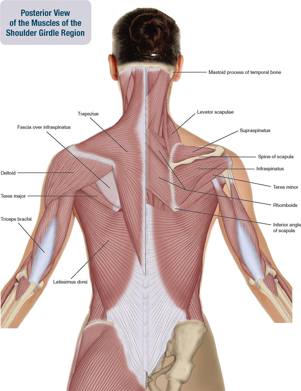
6 Muscles Of The Shoulder Girdle And Arm Musculoskeletal Key from musculoskeletalkey.com Normal anatomy, variants and checklist. Human anatomical atlas of the shoulder : Robin smithuis and henk jan van der woude. Diagram identifying the two clavicles between the sternum and the upper limbs. by leave a reply cancel reply. These joints can get arthritic and. Master arm and shoulder anatomy by studying this diagram depicts shoulder skeletal anatomy with parts and labels. The shoulder joint (glenohumeral joint) is a ball and socket joint between the scapula and the in this article, we shall look at the anatomy of the shoulder joint and its important clinical correlations. Assessment | biopsychology | comparative | cognitive | developmental | language | individual differences | personality | philosophy | social | methods | statistics | clinical | educational | industrial | professional items | world psychology |.
These joints can get arthritic and.
Mr is the best imaging modality to examen patients with shoulder pain and instability. Then, draw the shoulder girdle on top of the ribcage. Did not undergo a x ray or. We added an horizontal menu at. Left subtendinous bursa of subscapularis. The shoulder is a complex combination of bones and joints where many muscles act to provide the widest range of motion numerous muscles help stabilize the three joints of the shoulder while giving it motion. Robin smithuis and henk jan van der woude. Radiologists primarily perform shoulder imaging to assess injuries within the shoulder joint. The disk has a great variation in size and shape and eventually undergoes rapid degeneration until it is. In this episode we'll go over the simple structure and the anatomical details of both the clavicle and scapula. Learn their origins/insertions, functions & exercises. Shoulder anatomy diagram / anatomy of the left shoulder order : Ligaments are soft tissue that holds bone to bone.
We added an horizontal menu at. Mr is the best imaging modality to examen patients with shoulder pain and instability. Bone, then ligaments of the joint capsule, with tendons and muscles on top. Then, draw the shoulder girdle on top of the ribcage. Start studying shoulder anatomy diagram.
Applied Anatomy Of The Shoulder from www.orthopaedicmedicineonline.com The shoulder is made up of three bones: Sechrest, md narrates an animated tutorial on the basic anatomy of the shoulder. 7 draw labelled diagram showing the relations of shoulder joint. The shoulder is one of the largest and most complex joints in the body. It is made up of ligaments. Month ago i fell on my left shoulder while on a bush walk, hard fall. The nerve and blood supply to the supraspinatus, infraspinatus, rhomboid and levator scapulae muscles is illustrated. The shoulder joint is formed where the humerus (upper arm bone) fits into the scapula.
These joints can get arthritic and.
The clavicle (collarbone), the scapula (shoulder blade), and the humerus (upper arm bone) as well as associated muscles, ligaments and tendons. Webmd's shoulder anatomy page provides an image of the parts of the shoulder and describes its function, shoulder problems, and more. The human shoulder is made up of three bones: Diagram identifying the two clavicles between the sternum and the upper limbs. by leave a reply cancel reply. Master arm and shoulder anatomy by studying this diagram depicts shoulder skeletal anatomy with parts and labels. They're the muscles you see when you roll up. Anatomynote.com found shoulder bone anatomy from plenty of anatomical pictures on the internet. We added an horizontal menu at. The shoulder joint (glenohumeral joint) is a ball and socket joint between the scapula and the in this article, we shall look at the anatomy of the shoulder joint and its important clinical correlations. It is made up of ligaments. Your email address will not be published. This is because the deltoids are what you would consider the major muscles of the shoulder anatomy; Left subtendinous bursa of subscapularis.